Ask Dr Iain-Luxating Patellas
What should I know about Luxating Patella’s?
Patellar luxation is a common problem in many small breeds, including our Griffons. It has an inherited tendency in these breeds. The patella is another name for the kneecap and luxation means that the kneecap is unstable and in a severe case will actually dislocate and sit outside of the groove at the end of the femur bone (trochlear groove).
Patella Anatomy 101…..
Typically there is a misalignment of the quadriceps (thigh) muscle, patella, patellar tendon and the tibial crest at the top of the tibia bone which causes the patella to luxate within or even out of the trochlear groove in which it normally sits at the end of the femur bone. The quadriceps muscle is responsible for extending the knee (stifle) joint when it contracts. The patella tendon connects the muscle to the top of the tibia at the tibial crest. Have a feel of your Griffon’s knee or your own, and the tibial crest is that hard narrow ridge of bone at the front of the tibia. The patella tendon is the one that you can tap when the knee is relaxed and flexed to get that knee-jerk reflex. It is on the inside of this tendon where it goes over the end of the femur’s trochlear groove where the patella sits, and as the knee extends and flexes the patella will ride up and down the groove. For the knee to work properly all these bits need to be in a straight alignment!
What causes patella luxation?
When the patella is luxating it is usually because the tibial crest will be deviated medially (towards the inside aspect of the knee) which in turn will pull the patella so that it luxates medially. There is a grading system used by vets to describe the various degrees of luxation. The higher the score the worse the knee is affected and the more likely the dog will have problems. The patella is more likely to pop in and out when the knee is flexed because it is then that the patella tendon is relaxed.
The Medial Patella Luxation Grading System, commonly used by vets is from Grade 0 (normally positioned patella) to 4. Grade 1, where the patella can usually only be partially luxated by a vet on examination but immediately returns to the groove is considered to be virtually a normal finding of no concern by many veterinarians when examining small breed dogs. Dogs with Grade 2, 3 or 4 luxating patellas, especially if both knees are affected, are certainly at risk of developing arthritis and gait abnormalities and many veterinarians would not recommend them for breeding purposes. An added complication is that patella grading can differ in the same dog at different times during their life e.g. during her season or pregnancy a bitch may have a higher grade than when not in season or pregnant, and veterinarians can differ in their assessment of the same dog.
Grade 0, Patella cannot be luxated. Normal.
Grade 1, Patella can be luxated but spontaneous luxation during normal movement rarely occurs. Manual luxation by the vet pushing on the patella medially (towards to inner aspect of the knee) may be possible during physical exam but patella will return to normal position when pressure is released. Joint flexion and extension are normal.
Grade 2, Patella may luxate with flexion of stifle joint and can be manually displaced with lateral to medial pressure during physical exam. The patella remains luxated until it is reduced by the examiner or spontaneously reduces when the animal extends its stifle.
Grade 3, Patella remains medially luxated most of the time but may be manually reduced with the stifle in extension. However, after manual reduction, flexion and extension of the stifle result in reluxation of the patella.
Grade 4, There may be an 80-90 degree medial rotation of the top of the tibia and the crest. The patella is permanently luxated and cannot be manually repositioned. The femoral trochlear groove is very shallow or absent and there are marked deformities of the femur and tibia.
What can we do if my dog’s patella is luxating?
This is simple. If the dog is frequently found holding the leg up, or skipping around with the leg held up then they need surgery. These dogs are usually at least grade 2 or 3 in the affected leg. Sometimes a dog will have grade 0 or 1 patellas and then suddenly have a higher grade and lameness following an incident! It might follow leaping up to catch a Frisbee, jumping from the back porch or off a show trolley and the patellar tendon is stretched on landing and now we have a little dog with problems. If rest and anti-inflammatories don’t work or the vet can palpate significant luxation then surgery is required. If you don’t have surgery done then the knee will become increasingly arthritic because the patella and trochlear groove are actually components of the knee joint, they have smooth cartilage surfaces and lubricant production. With time, any unstable joint loses lubricant production and the smooth cartilage articular surfaces become worn and bony proliferation occurs, in other words, the joint develops painful chronic arthritis.
Some dogs with grade 4 patellas will have a crouching or crab like gait rather than skipping. Again, surgery is required to improve their quality of life. Sometimes you’ll see a dog skipping and then you might notice that they stretch the leg out behind themselves and then start walking normally again. This is because many dogs learn that by extending their leg they will pop the patella back into the trochlear groove. This doesn’t mean they won’t benefit from surgery.
Following surgery or in cases where there is no lameness or other gait issues but there is some instability of the patella/s, most vets will recommend chondroprotectives such as cartrophen or pentosan injections &/or daily glucosamine and even fish oil or other fatty acid supplements added to the diet. These can slow the progression of arthritis and even reverse some of the changes and modulate inflammatory changes.
Which surgery do I choose?
The ideal surgery is where we correct the alignment of the tibial crest, i.e. tibial crest transposition. The vet will use a fine saw (e.g. a sterile Junior hacksaw blade) to virtually cut the tibial crest off the front of the tibia bone. We try to cut the crest enough so that it can be pushed into a straight alignment with the trochlear groove and then use fine pins to reattach it to the tibia. Of course, this is done under a general anaesthetic! We will often deepen the trochlear groove, which often will become shallow if the patella hasn’t been sitting properly in it. The patient will go home with pain relief / anti-inflammatory medication and perhaps antibiotics. Usually a week or so later we will start a chondroprotective as discussed earlier. Sometimes, the small metal pins (also called K-wires) will migrate over time and possibly start irritating the muscles at the back or create swellings at the front of the tibia depending on the direction they have moved. If this happens then a quick operation to remove them can be done, because after several weeks the tibial crest will have healed and set in its new and correct position.
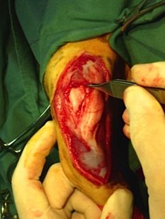
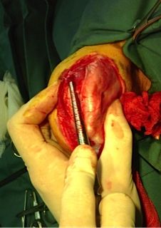
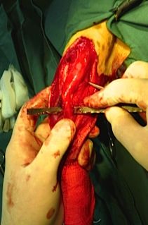
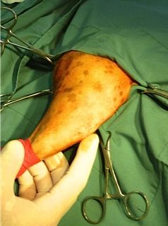
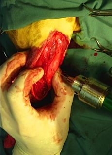
The right hind trochlea is exposed with The trochlear groove is being The crest has been cut from the tibia
the patellar tendon containing the patella deepened by using a rasp by 3cm. The patellar tendon overlies
Is pushed further to the right (medially) the tibial crest to which it is attached.
The crest is loose enough to be pushed
laterally (left) to a correct alignment.
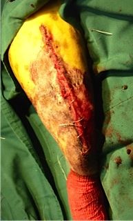
The first pin is being inserted through the Both pins have been inserted and cut,
now correctly aligned crest into the tibia bone. the joint capsule, fascia, subcutaneous tissue
and skin have been sutured and surgery is now completed.
Sometimes with a milder degree of medial luxation and if cost of surgery is a concern then a less expensive surgery might be successful. In these cases the vet might deepen the trochlear groove and place a non-absorbable suture around the patella and anchor this suture around a small bone at the back of the femur on the lateral side. This ‘patella de-rotational’ suture is in effect pulling the patella back into the groove. At the same time we might incise the tissue on medial aspect of the patella to lessen the tension there while excising some tissue and re-suturing the cut edges on the lateral side of the patella to aid pulling the patella into the trochlear groove.
In summary, don’t breed dogs with especially bilateral medially luxating Grade 2, 3 or 4 patellas, don’t throw Frisbees and don’t leave dogs unattended on top of show trolleys! If surgery is indicated, try to do the tibial crest transposition operation and ongoing chondroprotectives. If your vet doesn’t feel surgery is warranted or there are other reasons that may make surgery a higher than normal risk, then at least start chondroprotectives and weight reduction if necessary.
Iain Mitchell B.V.Sc(Hons), M.A.N.Z.C.V.Sc.
Contact Details
President: Mrs Colleen De Haan [email protected]
Secretary -Mrs Robin Simpson [email protected]